I had my first bone marrow biopsy over a year ago, just after being diagnosed with Hodgkin’s lymphoma. In preparation for my upcoming stem cell transplant, another biopsy was called for. This time I managed to get some photographs of the procedure, thanks to the assistance of my wife, Tammy. The biopsy procedure took place on the morning of Friday, July 1st.
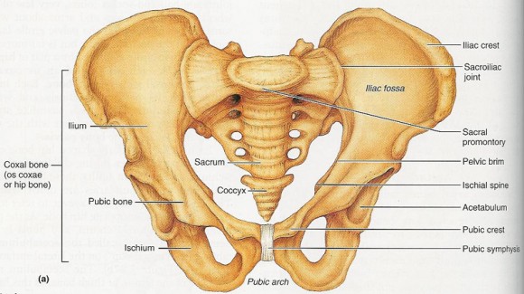
Both biopsies were taken from a location called the iliac crest. This is the top edge of the ilium; the largest bone in the pelvis.
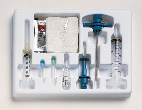
The procedure for taking a sample of bone marrow is not uncommon. There is a “kit” which is commonly used for this. It contains the necessary tools in a plastic tray.
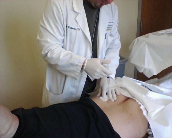
The iliac crest is easily accessed from either side of the spine. The area was cleaned with an antiseptic soap, and then the fun began. The first step was to numb the area with a local anesthetic; lidocaine, in this case.
He filled the syringe twice, emptying the bottle of lidocaine. He would inject a bit, move the needle further in, then inject some more. My skin was puffed up there because there was so much of it. The lidocaine causes a bit of a burning sensation at first, but does a great job of numbing the surrounding tissue.
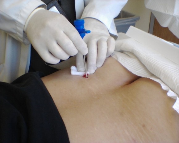
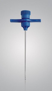 The lidocaine was given a minute or two to work its magic. It doesn’t take very long. A very narrow, deep incision was then made with a scalpel. I didn’t feel the incision being made at all. The incision is to make way for the Jamshidi® bone marrow biopsy aspiration needle.
The Jamshidi® biopsy needle has a razor-sharp beveled tip that is about 3mm across (just less than 1/8th inch). The needle itself is hollow and is slightly tapered to allow samples of bone marrow to be extracted easily. The handle makes it seem like they’re using a corkscrew on you. I guess that’s almost how the procedure looks, too. With the numbing of the local anesthetic, I only felt a little bit of pressure as it punctured the bone.
This photo shows the biopsy needle being inserted into the incision.
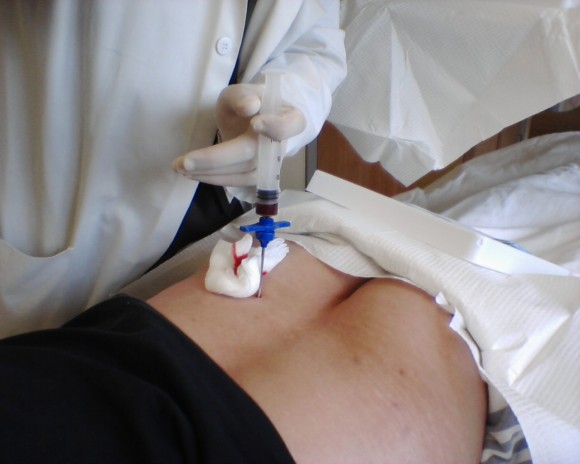
Once the biopsy needle is in place, it’s time to withdraw some tissue. The sample of spongy marrow is extracted through the needle with the attached insert. I never felt it. The marrow sample, about one inch (2.5cm) long, was pink, spongy, and sticky. Some was smeared onto slides, and the remainder was placed in a vial.
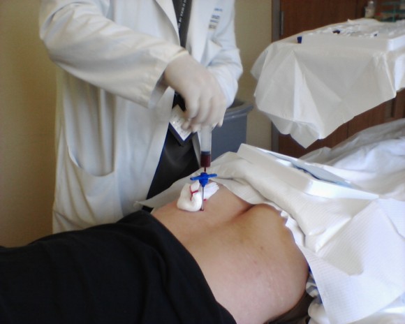
After the solid marrow sample is removed, liquid is drawn out by a syringe which attaches to the top of the biopsy needle.
The nurse who was performing the procedure had explained the steps before we started, and again as we went along. He had warned about this step as it would really “cramp up”, as far as pain went. He wasn’t kidding. It’s not easy to describe the feeling of the marrow being sucked out of your bones, but it isn’t really pleasant.
There really hadn’t been enough to aspirate when I had the first biopsy done. There was plenty of good marrow and fluid this time. I definitely felt it being extracted, too. It wasn’t enough to bring tears to my eyes, but I did say, “Shit!” when he was done. Thankfully, it only took a few seconds.
I’ll be back to see those guys again. I’ll be scheduled for another bone marrow biopsy about one hundred days after my upcoming stem cell transplant. Maybe I’ll get video next time.

2 comments
Sounds like you kept your composure pretty well. Kudos to going through with it (not like there’s much choice).
This was a rather fascinating entry for the uninitiated. Thanks for posting!
Hope the treatment goes well.
Ugh, sounds familiar…